Sinus Pause — ECG Rhythm | Telemetric Pro
A transient failure of the SA node to fire, producing a pause in the cardiac rhythm typically lasting less than 3 seconds before normal sinus activity resumes.
| Rate | Variable (underlying rate with pauses) |
|---|---|
| Rhythm | Irregular (pause disrupts regularity) |
| P Waves | Upright, uniform when present |
| PR Interval | 0.12–0.20 s (when conducted) |
| QRS Duration | < 0.12 s |
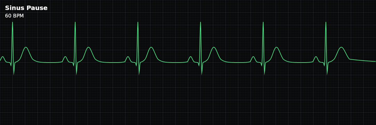
A sinus pause is a gap in the rhythm — the SA node temporarily fails to fire, and for a moment, there is nothing. No P wave, no QRS, no T wave. Just an unexpectedly long stretch of baseline. Then the SA node wakes up and the rhythm resumes as if nothing happened.
For monitor technicians, sinus pauses demand attention because the interpretation depends entirely on context. A 1.5-second pause during sleep is probably normal vagal tone. A 2.8-second pause while the patient is awake and symptomatic is a different story. The rhythm looks simple, but the clinical significance depends on duration, frequency, and patient symptoms.
What Changed from Normal Sinus Rhythm
Sinus pause changes one thing from NSR: the continuity of the rhythm. Between pauses, everything is normal — upright P waves, constant PR, narrow QRS. The pause itself is silence: an empty stretch of isoelectric baseline where a P-QRS-T complex should have been.
Five Criteria: Sinus Pause vs NSR
- Rate: Variable (effectively bradycardic)
- Between pauses, the rate may be normal (60-100). But the pauses lower the effective rate. If pauses are frequent or prolonged, the overall rate drops into the bradycardic range.
- Regularity: Irregular (pauses interrupt a regular rhythm)
- The underlying rhythm is regular sinus, but pauses create sudden gaps. The irregularity is not cyclic (like sinus arrhythmia) — it is abrupt and unpredictable.
- P Waves: Normal when present, absent during pause
- Before and after the pause, P waves are upright and uniform — the SA node is the pacemaker. During the pause, no P waves appear because the SA node is not firing.
- PR Interval: Normal when present (0.12-0.20s)
- PR intervals are constant in the conducted beats. There is no PR interval during the pause because there is no P wave.
- QRS Complex: Narrow (<0.12s)
- Ventricular conduction is normal in all conducted beats. QRS morphology does not change.
What a Sinus Pause Looks Like on the Strip
On the strip, you see a stretch of regular sinus rhythm, then a sudden gap — an empty baseline with no waveforms — followed by resumption of the sinus rhythm. The pause typically lasts less than 3 seconds. The beats before and after the pause look identical: same P waves, same PR, same QRS.
Sinus Pause vs SA Exit Block
Both produce pauses in an otherwise regular rhythm, but the mechanism is different: **Sinus Pause** — The SA node fails to generate an impulse. The pause is NOT an exact multiple of the P-P interval because the SA node's internal clock is reset. **SA Exit Block** — The SA node fires on schedule, but the impulse cannot exit the node to reach the atria. The pause IS an exact multiple of the P-P interval (usually 2x) because the SA node's clock keeps running.
Clinical Context for Monitor Technicians
Sinus pauses can be caused by increased vagal tone (common during sleep), medications that suppress the SA node (beta-blockers, calcium channel blockers, digoxin), intrinsic SA node disease (sick sinus syndrome), or electrolyte abnormalities. In hospitalized patients, medication effects are the most common reversible cause.
When to Escalate
**Notify promptly:** - Pause duration exceeds 3 seconds (approaches sinus arrest territory) - Patient reports symptoms during the pause — dizziness, lightheadedness, near-syncope - Pauses are frequent and recurring - An escape beat appears (junctional or ventricular) — this means the pause was long enough that a backup pacemaker had to fire **Document and monitor:** - Brief pauses (<2 seconds) during sleep with no symptoms - Isolated single pause in an otherwise stable patient
Medication-Related Pauses
Several drug classes suppress the SA node and can cause pauses. Beta-blockers (metoprolol, atenolol, carvedilol) slow automaticity and conduction. Calcium channel blockers (diltiazem, verapamil) have similar effects on the SA and AV nodes. Digoxin enhances vagal tone and can suppress SA node firing. Antiarrhythmics — particularly amiodarone and sotalol — have potent SA node depressant effects. In hospitalized patients, these medications are the most common reversible cause of pauses.
Timing matters. A 2.5-second pause that appears 30 minutes after an IV metoprolol push is a different clinical story than the same pause with no recent medication changes. The clinical team needs to correlate the pause with the medication administration record to decide whether to hold or adjust the dose.