Atrial Fibrillation (AFib) — ECG Rhythm | Telemetric Pro
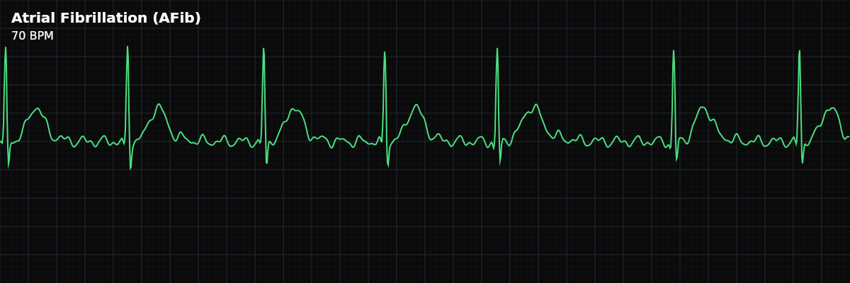
The most common sustained arrhythmia worldwide. Characterized by an irregularly irregular rhythm, absence of P-waves, and chaotic fibrillatory baseline activity.
| Rate | Variable (60–180 bpm ventricular) |
|---|---|
| Rhythm | Irregularly irregular |
| P Waves | Absent (fibrillatory baseline) |
| PR Interval | Not applicable |
| QRS Duration | < 0.12 s |
Every rhythm you have seen so far has been regular or close to it. Even when the rate was wrong — too fast in Sinus Tachycardia, too fast and locked in SVT — the spacing between beats was consistent. Atrial Fibrillation breaks that pattern completely. The R-R intervals are random, and no amount of measuring will reveal a repeating cycle. This "irregularly irregular" quality is not just a textbook phrase — it is the single most recognizable feature on a monitor strip.
AFib is also the most common sustained arrhythmia you will encounter in clinical practice. It appears across patient populations — post-surgical, cardiac, elderly, and sometimes in young patients with no known heart disease. Recognizing it quickly matters because the ventricular rate determines how urgently the clinical team needs to respond.
What Changed from Normal Sinus Rhythm
AFib is a **multi-criteria deviation** from NSR. The SA node is no longer driving the rhythm. Instead, hundreds of competing electrical circuits fire across the atria simultaneously. The AV node acts as a gatekeeper, allowing only some of these chaotic impulses to reach the ventricles — but it lets them through at random intervals, producing the irregularly irregular pattern you see on the strip.
Five Criteria: AFib vs Normal Sinus Rhythm
- Rate: Variable, 60-180 bpm (NSR: 60-100)
- The ventricular rate depends on how many impulses the AV node lets through. Controlled AFib runs 60-100 bpm. AFib with Rapid Ventricular Response (RVR) exceeds 100 bpm and often reaches 140-180.
- Regularity: Irregularly irregular (NSR: regular)
- This is the defining feature. Every R-R interval is different, and there is no repeating pattern — not even a roughly regular one. Use calipers on a 6-second strip and you will find no two intervals match.
- P Waves: Absent, replaced by f-waves (NSR: upright, one per QRS)
- There are no discrete P waves because the atria are not depolarizing in an organized way. The baseline between QRS complexes shows chaotic f-wave oscillations instead.
- PR Interval: Not applicable (NSR: 120-200ms)
- With no identifiable P wave, the PR interval cannot be measured. QRS complexes follow f-waves at random intervals.
- QRS Width: Narrow, <120ms (same as NSR)
- Despite the chaotic atrial activity, the ventricles still conduct normally through the His-Purkinje system. The narrow QRS confirms supraventricular origin.
Rate
Unlike other rhythms where "rate" is a single number, AFib has two rates: the **atrial rate** (350-600 bpm, unmeasurable on the strip) and the **ventricular rate** (what you actually see and report). The ventricular rate depends on how many impulses the AV node allows through, which is influenced by medications and the patient's autonomic nervous system.
For monitor technicians, the ventricular rate determines the clinical label: - **Controlled AFib** — Ventricular rate below 100 bpm at rest. The patient is on rate-control medication and the rhythm is being managed. - **AFib with RVR** (Rapid Ventricular Response) — Ventricular rate above 100 bpm. This is the version that triggers alarms and requires prompt notification.
Regularity
This is the feature that identifies AFib at a glance. Place your calipers at any R-R interval and walk them across the strip — they will never land in the same spot twice. The randomness is complete. There is no grouping, no pattern, no subtle regularity hiding underneath. This distinguishes AFib from rhythms that are merely "irregular": - **Sinus Arrhythmia** — Varies with breathing in a predictable cycle - **Atrial Flutter with variable block** — R-R intervals are multiples of a base interval - **Frequent PACs** — An underlying regular rhythm disrupted by early beats
P Waves and Fibrillatory Waves
In AFib, organized P waves are completely absent. The SA node is not driving atrial depolarization — hundreds of competing circuits are firing simultaneously. What you see on the baseline between QRS complexes is chaotic oscillation: **f-waves**.
f-waves come in two forms: **Coarse fibrillation** — Large, easily visible undulations on the baseline (amplitude >1mm). The chaotic activity is prominent and unmistakable. Coarse f-waves are more common in newer-onset AFib. **Fine fibrillation** — Low-amplitude oscillations that make the baseline appear nearly flat. Fine f-waves can be subtle enough to miss if you are not looking carefully. This pattern is more common in longstanding AFib where the atrial tissue has remodeled.