Normal Sinus Rhythm (NSR) — ECG Rhythm | Telemetric Pro
The baseline rhythm all others are measured against. Master NSR first — every abnormality is a departure from it.
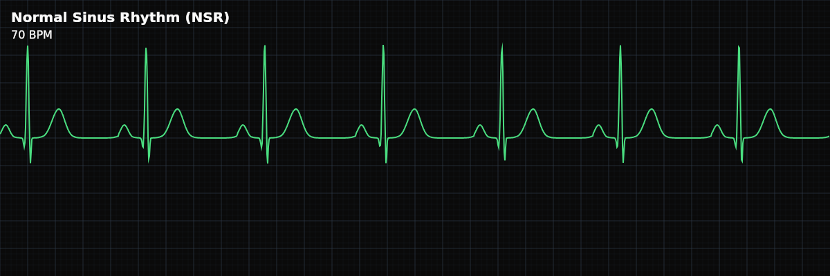
| Rate | 60–100 bpm |
|---|---|
| Rhythm | Regular |
| P Waves | Upright, one before each QRS |
| PR Interval | 0.12–0.20 s |
| QRS Duration | < 0.12 s |
Why This Rhythm Matters
Every rhythm you will learn in this curriculum is defined by how it *differs* from this one. Normal Sinus Rhythm is the reference standard — the rhythm produced when the heart's electrical system is functioning as designed, from origin to recovery. Before you can recognize what has gone wrong, you need a clear picture of what *right* looks like.
Think of NSR the way a musician thinks of a tuning fork. The tuning fork itself is not the performance — but without it, you cannot tell whether an instrument is sharp, flat, or in tune. NSR is your tuning fork for rhythm interpretation.
From Impulse to Ink: How the SA Node Builds a Heartbeat
The SA node — a cluster of specialized cells in the upper wall of the right atrium — fires an electrical impulse that spreads outward through the atrial tissue. That depolarization wave radiates from its origin like ripples expanding from where a stone enters water. As it sweeps through both atria, it produces the **P wave** on the tracing.
The impulse then reaches the AV node — the only electrical bridge between the atria and ventricles. Here, conduction deliberately slows. This brief pause (carried by slower calcium-channel conduction rather than the faster sodium channels used elsewhere) gives the atria time to finish contracting and push blood through the AV valves into the ventricles. On the tracing, this pause appears as the flat **PR segment** between the P wave and the QRS complex.
Once through the AV node, the impulse accelerates through the His bundle, splits into the right and left bundle branches, and fans out through the Purkinje network to depolarize the ventricular muscle nearly simultaneously. This rapid, coordinated ventricular depolarization produces the tall, narrow **QRS complex**. The ventricles then recover — ventricular repolarization inscribes the **T wave**. The strip that follows labels each step in this sequence.
This entire sequence — SA node fires, atria depolarize (P), AV node pauses (PR segment), ventricles depolarize (QRS), ventricles recover (T) — repeats with every heartbeat. Each cycle appears on the monitor as one **P-QRS-T complex**. In Normal Sinus Rhythm, this cycle repeats at a steady rate between 60 and 100 times per minute.
The Five Criteria: Your NSR Checklist
A systematic approach to rhythm analysis asks five questions of every strip. When all five answers fall within normal limits, the rhythm is NSR. When any answer deviates, you have a starting point for identifying *which* abnormality is present.
- Rate: 60–100 bpm
- Measured by the RR interval. At 60 bpm the interval is 1000 ms (5 large boxes); at 100 bpm it is 600 ms (3 large boxes). **Why it matters:** Outside this range the SA node isn't setting the pace — you're looking at a brady or tachy rhythm.
- Regularity: Regular
- RR intervals are equal throughout the tracing. The SA node fires at a constant rate, producing evenly spaced complexes. **Why it matters:** Irregular spacing is your first clue something else is firing — ectopy, block, or atrial chaos.
- P waves: Upright, uniform, one before every QRS
- Upright in Lead II (confirms top-down atrial depolarization from the SA node). Identical morphology beat to beat (confirms a single, consistent origin). Duration < 120 ms, amplitude < 2.5 mm. **Why it matters:** Each P wave proves the impulse started in the SA node and traveled the normal route.
- PR interval: 120–200 ms, constant
- Measured from the start of the P wave to the first deflection of the QRS. Represents atrial depolarization plus the AV node delay. The same duration in every complex. **Why it matters:** Too short means a shortcut exists; too long means the AV node is holding traffic.
- QRS complex: Narrow (< 120 ms), one after every P wave
- A narrow QRS confirms that ventricular depolarization followed the normal His-Purkinje pathway. Every P wave produces a QRS — no dropped beats. **Why it matters:** A wide QRS means the ventricles aren't using the highway — bundle branch problem or ventricular origin.
Rate
The SA node's intrinsic firing rate — shaped by the balance of sympathetic and parasympathetic tone — produces a heart rate between 60 and 100 bpm. That range defines NSR. Below 60, the same SA-driven mechanism is classified as Sinus Bradycardia; above 100, Sinus Tachycardia. The underlying process hasn't changed — only the speed.
Rate is measured by the **RR interval** — the distance between two consecutive R waves. At 60 bpm the RR interval is 1000 ms (25 mm / 5 large boxes). At 100 bpm it shrinks to 600 ms (15 mm / 3 large boxes). Any RR interval that falls between those two boundaries corresponds to a rate within the NSR range.