First Degree AV Block — ECG Rhythm | Telemetric Pro
A conduction delay at the AV node where every P wave is conducted to the ventricles, but the PR interval is prolonged beyond 200ms. Not a true block — more accurately a "first degree AV delay."
| Rate | 60–100 bpm |
|---|---|
| Rhythm | Regular |
| P Waves | Upright, one before each QRS |
| PR Interval | > 0.20 s (prolonged) |
| QRS Duration | < 0.12 s |
First Degree AV Block is the mildest of the AV conduction abnormalities. Every impulse from the SA node reaches the ventricles — nothing is blocked. The only abnormality is that it takes longer than normal for the impulse to cross the AV node. This delay shows up on the strip as a prolonged PR interval: greater than 200ms (one large box) instead of the normal 120-200ms.
For monitor technicians, first degree AV block is important to recognize because it sits at the beginning of the AV block spectrum. By itself, it is usually benign. But knowing the PR interval is prolonged is essential context — if the patient later develops progressive PR lengthening (Wenckebach) or dropped beats (Type II), you can identify the progression because you noticed the prolonged PR baseline.
What Changed from Normal Sinus Rhythm
First degree AV block changes exactly one thing from NSR: the PR interval is prolonged. Everything else — the rate, the rhythm, the P waves, the QRS — remains identical to normal sinus rhythm. This is why it is the easiest AV block to miss: if you do not measure the PR interval, the strip looks completely normal.
Five Criteria: First Degree AV Block vs NSR
- Rate: Normal (depends on underlying rhythm)
- First degree block does not affect the heart rate. The rate depends entirely on the underlying sinus rhythm (typically 60-100 BPM).
- Regularity: Regular
- RR intervals are constant. The rhythm is identical to NSR in every respect except the PR interval.
- P Waves: Normal, upright, one per QRS
- Every P wave is followed by a QRS. P-wave morphology is normal. 1:1 AV conduction is fully preserved — no beats are dropped.
- PR Interval: Prolonged (>200ms) and constant
- Greater than one large box on standard paper. The prolongation is the same from beat to beat — the PR does not progressively lengthen (which would be Wenckebach).
- QRS Complex: Narrow (<0.12s)
- The block is at the AV node level, above the bundle branches. Ventricular conduction is completely normal.
What First Degree AV Block Looks Like on the Strip
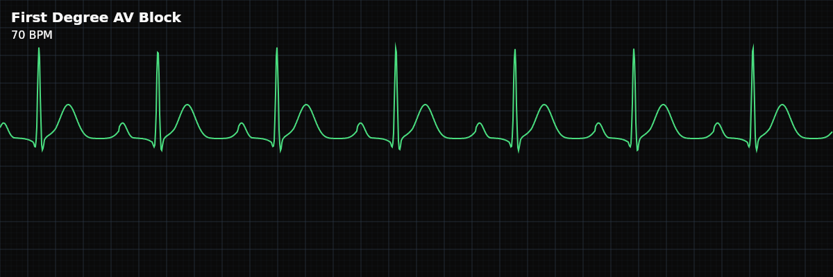
On the strip, first degree AV block looks like normal sinus rhythm with an unusually long gap between the P wave and the QRS complex. The P wave ends, and there is a noticeable pause before the QRS begins. This gap is the prolonged PR interval. If you measure it, it exceeds 200ms (one large box). Everything else about the strip looks completely normal.
The AV Block Spectrum
First degree AV block is the mildest in a spectrum of AV conduction abnormalities: **First Degree** — PR prolonged, every beat conducted. A delay, not a true block. **Second Degree Type I (Wenckebach)** — PR progressively lengthens until a beat is dropped. Pattern repeats. **Second Degree Type II (Mobitz II)** — PR is constant, but beats drop without warning. More concerning. **Third Degree (Complete)** — No atrial impulses conduct to the ventricles. Complete dissociation. Understanding this spectrum helps you recognize progression: if a patient with known first degree block starts showing progressive PR lengthening, they may be developing Wenckebach.
Clinical Context for Monitor Technicians
First degree AV block is common and usually benign. It can be caused by medications that slow AV conduction (beta-blockers, calcium channel blockers, digoxin, amiodarone), increased vagal tone (athletes, during sleep), age-related fibrosis of the conduction system, inferior MI affecting the AV node, myocarditis, or electrolyte abnormalities (hyperkalemia).
When to Escalate
**Notify promptly:** - New first degree AV block (not previously documented for this patient) - PR interval is markedly prolonged (>300ms) - PR is progressively lengthening (may be developing Wenckebach) - Dropped beats appear (progression to second degree block) - Patient is symptomatic (rare with first degree alone, but possible at extreme PR intervals) **Document and monitor:** - Known, stable first degree AV block with no change in PR interval - First degree block in a patient on AV-nodal blocking medications (expected finding)
Putting It Together
First degree AV block is a prolonged but constant PR interval (>200ms) with 1:1 conduction — every P wave produces a QRS. It is not a true block but a conduction delay. Usually benign, it can be caused by medications, vagal tone, or aging. The clinical importance is recognizing it as a baseline and watching for progression to higher-degree blocks.