Sinus Arrhythmia — ECG Rhythm | Telemetric Pro
A normal sinus rhythm variant where the heart rate cyclically increases during inspiration and decreases during expiration, reflecting healthy autonomic modulation.
| Rate | 60–100 bpm |
|---|---|
| Rhythm | Irregular (varies with respiration) |
| P Waves | Upright, uniform, one before each QRS |
| PR Interval | 0.12–0.20 s |
| QRS Duration | < 0.12 s |
Sinus Arrhythmia is one of the first rhythms that can fool a new monitor technician into thinking something is wrong. You see an irregular rhythm on the strip, and irregular usually means trouble. But sinus arrhythmia is the exception — it is a completely normal physiological variant that reflects a healthy heart with intact autonomic control.
The key to recognizing sinus arrhythmia is understanding what makes it different from pathological irregularity. In AFib, the irregularity is chaotic — no two RR intervals follow a pattern. In sinus arrhythmia, the irregularity is cyclic: the heart speeds up when the patient breathes in and slows down when they breathe out. This respiratory pattern is the hallmark, and once you learn to see it, the rhythm is unmistakable.
What Changed from Normal Sinus Rhythm
Sinus arrhythmia changes exactly one thing from NSR: the regularity of the RR intervals. Everything else — the P-wave morphology, the PR interval, the QRS complex, the conduction pathway — remains identical. The SA node is still in control; it is simply varying its firing rate with respiration.
Five Criteria: Sinus Arrhythmia vs NSR
- Rate: 60-100 BPM (average)
- The average rate stays in the normal sinus range. The rate rises slightly during inspiration and falls during expiration, but the overall average is 60-100.
- Regularity: Regularly Irregular (cyclic)
- RR intervals vary by more than 120ms, but in a predictable, phasic pattern that follows the breathing cycle. This cyclic pattern is the defining feature.
- P Waves: Normal, Uniform
- Every P wave looks the same — upright, rounded, one per QRS. The SA node is the pacemaker throughout. This confirms the rhythm has not shifted to another focus.
- PR Interval: Constant (0.12-0.20s)
- The PR interval does not change despite the varying rate. This is a critical distinction from Wandering Atrial Pacemaker (WAP), where the PR varies because the pacemaker focus shifts.
- QRS Complex: Narrow (<0.12s)
- Ventricular conduction is completely normal. The QRS morphology does not change.
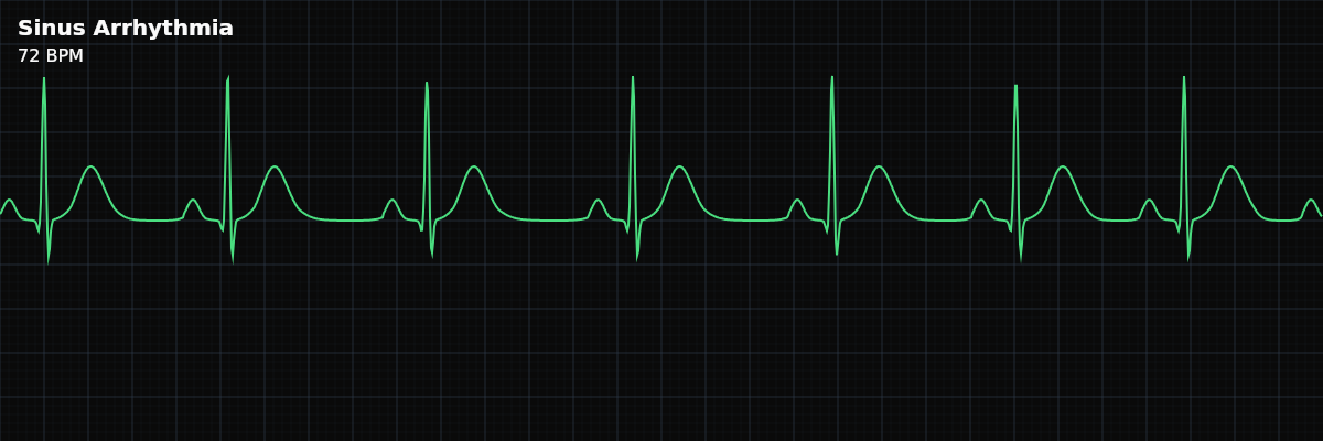
What Sinus Arrhythmia Looks Like on the Strip
On the strip, sinus arrhythmia looks almost identical to NSR — the same upright P waves, the same narrow QRS complexes, the same T waves. The only difference is the spacing: complexes cluster slightly closer together during inspiration and spread apart during expiration. The variation is gradual, not sudden — there are no abrupt pauses or premature beats.
Sinus Arrhythmia vs Wandering Atrial Pacemaker (WAP)
Both sinus arrhythmia and WAP are benign irregular rhythms at rates under 100 bpm. The P-wave morphology is the deciding factor: | Feature | Sinus Arrhythmia | WAP | |---------|-------------------|-----| | P-wave morphology | **Constant** (same shape every beat) | **Variable** (≥ 3 distinct shapes) | | PR interval | **Constant** | **Variable** (changes with P morphology) | | Rate variation | Phasic, linked to respiration | Gradual, not respiratory | | Mechanism | Vagal modulation of SA node rate | Shifting pacemaker site among atria/AV junction | If the P waves all look the same and the PR interval is constant, the pacemaker has not moved — you are looking at sinus arrhythmia. If the P-wave shape shifts from beat to beat and the PR varies with it, the origin is migrating — that is WAP.
Sinus Arrhythmia vs Atrial Fibrillation
This is the distinction that matters most on the floor. Both rhythms are irregular, but they are irregular in completely different ways: **Sinus Arrhythmia** — Cyclic, predictable irregularity. P waves are present and uniform. Baseline is clean and isoelectric. PR intervals are constant. The patient is usually young and healthy. **Atrial Fibrillation** — Chaotic, unpredictable irregularity. P waves are absent, replaced by fibrillatory waves. Baseline is wavy and disorganized. No measurable PR interval. Often seen in older patients or those with heart disease.
Clinical Context for Monitor Technicians
Sinus arrhythmia is most prominent in children, young adults, and athletes — populations with strong vagal tone. It tends to diminish with age as autonomic responsiveness decreases. Seeing sinus arrhythmia on a young patient is actually a sign of healthy cardiac autonomic function.
When to Escalate
Standard respiratory sinus arrhythmia does not require escalation. However, escalate if: - The irregularity is chaotic rather than cyclic (may be AFib, not sinus arrhythmia) - P-wave morphology changes between beats (may be Wandering Atrial Pacemaker) - The variation does not correlate with breathing in an elderly patient (non-respiratory type — possible SA node disease) - The patient reports symptoms such as dizziness or palpitations during the irregular phase