Right Bundle Branch Block (RBBB) — ECG Rhythm | Telemetric Pro
A conduction abnormality where the right bundle branch is blocked, forcing the right ventricle to depolarize via muscle-to-muscle conduction from the left ventricle. The QRS is wide (>= 0.12s) with a characteristic RSR’ (M-shape) pattern in V1.
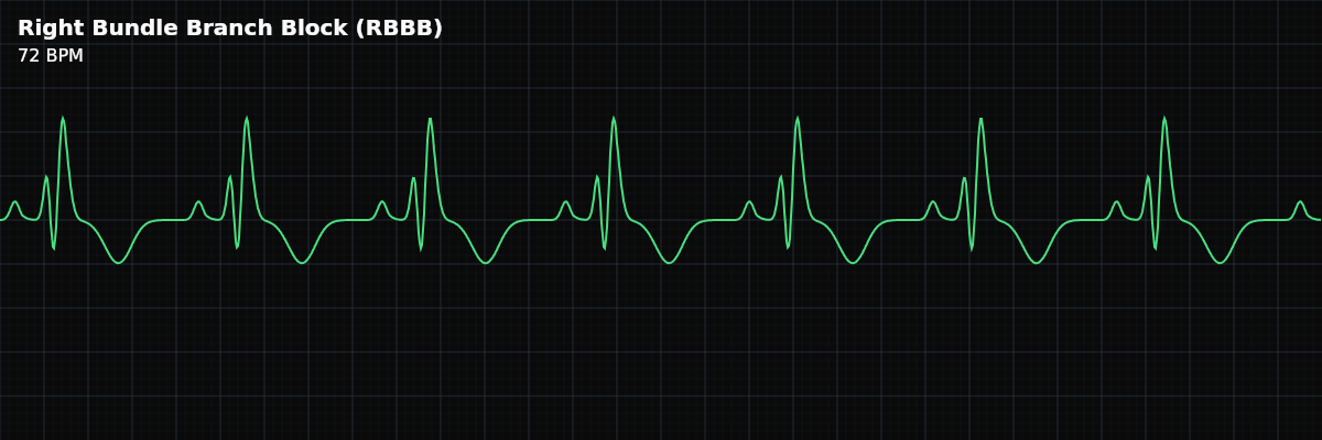
| Rate | Depends on underlying rhythm |
|---|---|
| Rhythm | Regular (usually sinus) |
| P Waves | Normal (sinus origin) |
| PR Interval | 0.12–0.20 s (normal) |
| QRS Duration | ≥ 0.12 s (wide) |
| V1 Pattern | RSR’ (M-shape) |
| V6 Pattern | Wide S wave |
Right Bundle Branch Block is a conduction abnormality, not a rhythm disturbance. The SA node fires normally, the atria depolarize normally, and the AV node conducts normally. The problem is downstream: the right bundle branch is blocked, so the electrical impulse cannot travel its usual fast path to the right ventricle. Instead, the right ventricle depolarizes late, activated by slow muscle-to-muscle spread from the left side. This delayed activation widens the QRS and creates a distinctive pattern on the monitor.
For monitor technicians, recognizing RBBB matters for two reasons. First, a wide QRS can mimic ventricular rhythms — you need to distinguish RBBB from ventricular tachycardia (VT). Second, the clinical significance depends entirely on context: chronic, documented RBBB is a stable baseline finding, while new-onset RBBB may signal an acute process like pulmonary embolism (PE) or right ventricular strain.
At a Glance
What Changed from Normal Sinus Rhythm
In normal sinus rhythm, the impulse travels down both bundle branches simultaneously. Both ventricles activate at nearly the same time, producing a narrow QRS (< 0.12 seconds). In RBBB, the right bundle is blocked. The left ventricle still activates on schedule, but the right ventricle must wait for the wave of depolarization to spread across from the left side, muscle cell to muscle cell. This slow detour widens the QRS to 0.12 seconds or more and creates the characteristic terminal deflection seen in the right-sided leads.
Five Criteria: RBBB vs Normal Sinus Rhythm
- Rate: Depends on underlying rhythm
- RBBB does not affect the heart rate. The rate is determined by the underlying sinus rhythm (typically 60-100 BPM). You can have RBBB with sinus bradycardia, normal sinus rhythm, or sinus tachycardia.
- Regularity: Regular (usually)
- The rhythm is regular because the underlying sinus mechanism is unchanged. RBBB can coexist with irregular rhythms (AFib with RBBB, for example), but the block itself does not cause irregularity.
- P Waves: Normal, upright, one per QRS
- P waves are present and normal because atrial depolarization is unaffected. The SA node fires normally, and AV conduction is intact. Every P wave is followed by a (widened) QRS.
- PR Interval: Normal (0.12-0.20s)
- The PR interval is normal because AV node conduction is not affected by a bundle branch block. The delay occurs below the AV node, in the ventricular conduction system.
- QRS Duration: Wide (>= 0.12s)
- This is the defining feature. The QRS is 0.12 seconds or wider (3+ small boxes). The extra width comes from the delayed right ventricular activation appended to the end of the QRS complex.
V1 Pattern: RSR’ (M-Shape)
The hallmark of RBBB is the RSR’ pattern in lead V1 (a right-sided chest lead). This pattern looks like the letter M: **R** — The initial small upward deflection represents normal septal depolarization (left to right). **S** — The downward deflection represents left ventricular activation (the normal, on-time part). **R’** — The second upward deflection represents the delayed right ventricular activation. This is the extra piece that widens the QRS. Because V1 sits over the right ventricle, this late rightward force is directed toward V1 and produces a positive (upward) deflection.
V6 Pattern: Wide S Wave
In V6 (a left-sided lead), RBBB produces a wide, slurred S wave following the R wave. The R wave is the normal left ventricular activation coming toward V6. The wide S wave is the delayed right ventricular activation moving away from V6 (toward the right side). This is the mirror image of what V1 shows — same late right ventricular activation, but seen from the opposite side of the chest.
The MaRRoW Mnemonic
Vocabulary: **MaRRoW** — A memory aid for bundle branch block patterns: **Ma** — In **M**arching order: V1 first, then V6. **RR** — **R**BBB has an **R**SR’ (M-shape) in V1. Two R’s = two bumps = M. **W** — The opposite lead (V6) shows a **W**ide S wave, like a W-shape. So RBBB = M in V1, W in V6. LBBB is the opposite: W in V1, M in V6.
RBBB vs LBBB vs Normal Sinus Rhythm
**Key differences at a glance:** **NSR** — Narrow QRS. Both ventricles activated simultaneously via intact bundle branches. **RBBB** — Wide QRS. Terminal R’ (upward) in V1; wide S wave in V6. Right ventricle late. **LBBB** — Wide QRS. Deep QS or rS (downward) in V1; broad, notched R in V6. Left ventricle late.
Clinical Context for Monitor Technicians
RBBB can be found in healthy individuals with no heart disease — it is sometimes an incidental finding. It can also result from right ventricular strain, pulmonary embolism (PE), right heart enlargement (cor pulmonale), coronary artery disease, myocarditis, or congenital heart defects (especially atrial septal defect).
**Chronic vs new-onset:** **Chronic RBBB** — Documented in the patient’s chart, present on prior ECGs. A stable baseline finding that requires no acute intervention. Document it as part of the patient’s rhythm description. **New-onset RBBB** — Not previously documented. This is clinically significant and warrants prompt notification. New RBBB can indicate acute right ventricular strain (pulmonary embolism), progression of conduction system disease, or acute coronary syndrome.