Premature Ventricular Contractions (PVCs) — ECG Rhythm | Telemetric Pro
Early beats originating from an ectopic focus in the ventricles. PVCs are wide, bizarre-looking QRS complexes that appear before the next expected sinus beat, typically followed by a compensatory pause. They are the most common ventricular ectopy.
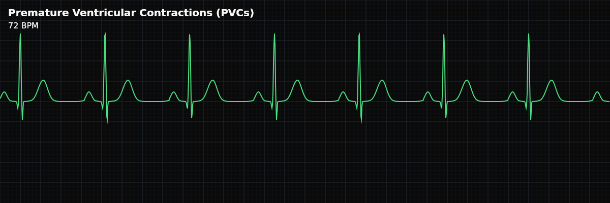
Premature Ventricular Contractions are the most common type of ventricular ectopy. A PVC occurs when an irritable focus somewhere in the ventricular myocardium fires before the next expected sinus beat. Because the impulse originates in the ventricle rather than traveling through the normal His-Purkinje system, the resulting QRS is wide (>= 0.12 seconds) and has an abnormal, often bizarre morphology. The PVC is typically followed by a compensatory pause before the next sinus beat.
For monitor technicians, PVCs are one of the most frequently seen findings on a telemetry monitor. Most PVCs are benign — they occur in healthy hearts and are often triggered by caffeine, stress, or electrolyte shifts. But PVCs in certain patterns, frequencies, or clinical contexts can be precursors to dangerous ventricular arrhythmias. Knowing which PVC patterns require escalation is a core monitoring skill.
What a PVC Looks Like on the Strip
A PVC has four identifying features on the strip: **1. It is early** — The PVC fires before the next expected sinus beat, interrupting the regular rhythm. **2. The QRS is wide (>= 0.12s)** — Because the impulse starts in ventricular muscle and spreads cell-to-cell rather than through the fast conduction system, the QRS is wide and often bizarre-looking. **3. No preceding P wave** — The beat originates in the ventricle, not from atrial depolarization. There is no P wave before the wide QRS. (Sometimes a sinus P wave is buried in the PVC, but it is coincidental — it did not cause the PVC.) **4. Compensatory pause** — After the PVC, there is usually a pause before the next sinus beat. This full compensatory pause occurs because the sinus node is not reset by the PVC — it continues firing on schedule, and the next sinus beat arrives on time.
Why PVCs Look Wide
Normal sinus beats travel through the His-Purkinje system — a high-speed electrical highway that activates both ventricles nearly simultaneously in under 0.12 seconds. A PVC bypasses this system entirely. Starting from a focus somewhere in the ventricular muscle, the impulse must crawl through myocardial tissue cell by cell. This is much slower, which is why the QRS is wide (>= 0.12 seconds) and often looks distorted or bizarre compared to the narrow sinus QRS.
Unifocal vs Multifocal PVCs
**Unifocal PVCs** — All PVCs look the same on the strip. They originate from the same ventricular focus, so every PVC has an identical morphology (shape, width, direction). Unifocal PVCs are generally more benign because they come from a single irritable spot. **Multifocal PVCs** — PVCs have different shapes on the strip. They originate from different ventricular foci, so the QRS morphology varies from one PVC to the next. Multifocal PVCs are more concerning because they indicate widespread ventricular irritability — multiple sites in the ventricle are firing inappropriately.
PVC Patterns
PVCs can occur in recognizable patterns that carry different clinical implications:
- Isolated PVCs
- Single PVCs scattered among normal beats with no predictable pattern. The most common and usually most benign form. Occasional isolated PVCs are a normal finding.
- Bigeminy
- Every other beat is a PVC: normal-PVC-normal-PVC. A repeating pattern of one normal beat alternating with one PVC. Common and often benign, but sustained bigeminy can reduce cardiac output because PVCs typically eject less blood than normal beats.
- Trigeminy
- Every third beat is a PVC: normal-normal-PVC-normal-normal-PVC. A repeating pattern with two normal beats between each PVC.
- Couplet
- Two consecutive PVCs in a row. More concerning than isolated PVCs because it indicates the ventricular focus fired twice in rapid succession. Couplets suggest increased ventricular irritability.
- Triplet / Non-Sustained VT (NSVT)
- Three consecutive PVCs. By definition, three or more consecutive ventricular beats constitute ventricular tachycardia. If it lasts less than 30 seconds and terminates spontaneously, it is called non-sustained VT (NSVT). This always requires notification.
R-on-T Phenomenon
The R-on-T phenomenon occurs when a PVC lands on the T wave of the preceding beat. The T wave represents ventricular repolarization — a vulnerable period when the heart is resetting its electrical state. A PVC firing during this vulnerable period can trigger ventricular fibrillation (VFib). On the strip, R-on-T looks like a PVC that falls directly on top of the preceding T wave, rather than appearing after the T wave has completed. The PVC seems to start before the T wave ends.