Premature Atrial Contractions (PACs) — ECG Rhythm | Telemetric Pro
Early beats originating from an ectopic focus in the atria. PACs produce a P wave with abnormal morphology (different shape from sinus P waves) followed by a narrow QRS complex, since conduction through the ventricles follows the normal pathway.
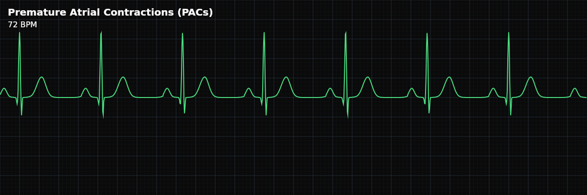
Premature Atrial Contractions are early beats that originate from an ectopic focus somewhere in the atria — anywhere except the sinoatrial (SA) node. The ectopic focus fires before the next expected sinus beat, producing a premature P wave with a different shape from the normal sinus P waves. Because the impulse still travels through the AV node and His-Purkinje system normally, the QRS that follows is narrow (< 0.12 seconds).
For monitor technicians, PACs are among the most common findings on a telemetry monitor. They are usually benign and require no intervention. The clinical importance of PACs lies in two areas: distinguishing them from PVCs (which have different clinical implications) and recognizing when frequent PACs may be triggering or foreshadowing sustained supraventricular arrhythmias like SVT or atrial fibrillation.
Identifying a PAC on the Strip
A PAC has four identifying features: **1. It is early** — The PAC appears before the next expected sinus beat, interrupting the regular RR interval. **2. The P wave looks different** — The P wave before the PAC has a different morphology (shape) from the normal sinus P waves. It may be taller, shorter, notched, inverted, or have a different contour. This difference occurs because the impulse originates from a different location in the atria than the SA node. **3. The QRS is narrow (< 0.12s)** — Unlike PVCs, PACs travel through the normal ventricular conduction system. The impulse enters the AV node, travels down the bundle branches, and activates both ventricles simultaneously via the His-Purkinje system. The resulting QRS looks identical to the patient’s normal sinus QRS. **4. Non-compensatory pause** — After the PAC, there is usually a pause, but it is shorter than the full compensatory pause seen with PVCs. The ectopic atrial impulse typically resets the sinus node, so the SA node restarts its cycle from the PAC rather than continuing on its original schedule.
What Frequent PACs Look Like
PAC vs PVC: The Critical Distinction
The fastest way to distinguish a PAC from a PVC is the QRS width: **Narrow QRS (< 0.12s) = PAC** — The impulse used the normal conduction system. It started above the ventricles (atria) and traveled the normal pathway down. **Wide QRS (>= 0.12s) = PVC** — The impulse originated in the ventricle and spread via slow muscle-to-muscle conduction, widening the QRS. There is one exception: an aberrantly conducted PAC. Occasionally, a very early PAC arrives at the bundle branches before they have fully repolarized, causing the QRS to conduct with a temporary bundle branch block pattern (usually RBBB). This aberrantly conducted PAC has a wide QRS but is preceded by an abnormal P wave — the P wave is the clue that it is atrial in origin despite the wide QRS.
Going Deeper — Ashman Phenomenon
When a PAC arrives early enough that one bundle branch has not fully recovered from the previous depolarization, the impulse conducts with a wide QRS that mimics a PVC. This is **aberrant ventricular conduction** — the atrial impulse is conducted, but one bundle branch is still refractory, so the QRS widens. Key features of PAC with aberrancy: 1. **Preceded by a premature P-wave** — look for subtle distortion of the preceding T-wave 2. **RBBB pattern is most common** — the right bundle branch has the longest refractory period, so it is the last to recover 3. **Initial QRS vector preserved** — the first few milliseconds of the QRS look the same as normal beats 4. **Incomplete compensatory pause** — same as a normally conducted PAC (SA node resets), unlike PVCs which typically have a full compensatory pause