Left Bundle Branch Block (LBBB) — ECG Rhythm | Telemetric Pro
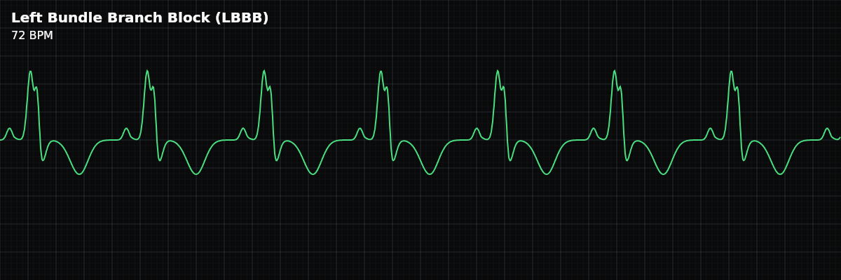
A conduction abnormality where the left bundle branch is blocked, forcing the left ventricle to depolarize via slow muscle-to-muscle conduction from the right ventricle. The QRS is wide (>= 0.12s, often >= 0.14s) with a QS or rS pattern in V1 and a broad, notched R wave in V6.
| Rate | Depends on underlying rhythm |
|---|---|
| Rhythm | Regular (usually sinus) |
| P Waves | Normal (sinus origin) |
| PR Interval | 0.12–0.20 s (normal) |
| QRS Duration | ≥ 0.12 s (wide, often ≥ 0.14 s) |
| V1 Pattern | QS or rS (deep negative) |
| V6 Pattern | Broad, notched R wave |
Left Bundle Branch Block is a conduction abnormality with higher clinical significance than its right-sided counterpart. Like RBBB, the SA node fires normally and AV conduction is intact — the block is in the ventricular conduction system. But because the left bundle branch serves the larger, thicker-walled left ventricle, LBBB has two important consequences that RBBB does not: it changes the expected direction of ST segments and T waves, and new-onset LBBB with ischemic symptoms is treated as a STEMI equivalent.
For monitor technicians, LBBB demands more attention than RBBB. You need to recognize the pattern, distinguish it from ventricular rhythms, understand that the ST/T-wave changes you see are expected (not necessarily ischemic), and know that a new LBBB in a symptomatic patient requires immediate escalation.
At a Glance
What Changed from Normal Sinus Rhythm
In NSR, both ventricles activate simultaneously via their bundle branches, producing a narrow QRS. In LBBB, the left bundle is blocked. The right ventricle activates on time, but the left ventricle depolarizes late — activated by slow cell-to-cell spread from the right side. Because the left ventricle is much larger, this delayed activation dominates the QRS, producing a wide, often notched complex. The altered depolarization pathway also changes repolarization, producing the ST/T-wave discordance that is characteristic of LBBB.
Five Criteria: LBBB vs Normal Sinus Rhythm
- Rate: Depends on underlying rhythm
- LBBB does not affect the heart rate. The rate is set by the underlying sinus rhythm. LBBB can coexist with bradycardia, normal rate, or tachycardia.
- Regularity: Regular (usually)
- The rhythm is regular when the underlying mechanism is sinus. LBBB can coexist with atrial fibrillation (AFib with LBBB) — the wide QRS makes it look alarming, but the irregularity comes from the AFib, not the block.
- P Waves: Normal, upright, one per QRS
- Atrial depolarization is unaffected. P waves are present and normal in sinus rhythm with LBBB. Every P wave is followed by a (widened) QRS.
- PR Interval: Normal (0.12-0.20s)
- AV node conduction is intact. The delay occurs below the AV node, in the left bundle branch. A prolonged PR with LBBB suggests coexisting first degree AV block.
- QRS Duration: Wide (>= 0.12s, often >= 0.14s)
- The QRS is wide because the left ventricle activates late via slow muscle-to-muscle conduction. LBBB often produces a wider QRS than RBBB because the left ventricle is larger and takes longer to depolarize via this indirect path.
V1 Pattern: QS or rS (Deep Negative Deflection)
In lead V1 (a right-sided chest lead), LBBB produces a predominantly negative deflection: **QS pattern** — The entire QRS is negative (downward). No initial R wave at all. This is the most common V1 pattern in LBBB. **rS pattern** — A tiny initial r wave (small upward blip from septal depolarization) followed by a deep S wave. The S wave dominates. Why? In LBBB, the normal left-to-right septal depolarization is lost or reversed. The dominant electrical force moves away from V1 (toward the left ventricle), producing the deep negative deflection. This is the opposite of RBBB, where V1 shows a terminal positive deflection.
V6 Pattern: Broad, Notched R Wave
In V6 (a left-sided lead), LBBB produces a tall, broad R wave that is often notched or has a plateau at its peak. The R wave is tall because the delayed left ventricular activation moves toward V6. The notch occurs because the right and left ventricles activate sequentially rather than simultaneously — you see the right ventricle’s contribution, then a brief hitch, then the dominant left ventricular activation. There is typically no Q wave in V6 (the normal septal Q is absent because septal depolarization is reversed in LBBB). The absence of the septal Q wave in V6 is itself a diagnostic clue.
The WiLLiaM Mnemonic
Vocabulary: **WiLLiaM** — A memory aid for LBBB patterns (paired with MaRRoW for RBBB): **W** — V1 shows a **W**-shape: QS or rS pattern (deep negative deflection). **LL** — This is **L**eft bundle branch block — **L**BBB. **M** — V6 shows an **M**-shape: broad, notched R wave (positive deflection). So LBBB = W in V1, M in V6. RBBB is the opposite (MaRRoW): M in V1, W in V6.
ST/T-Wave Changes: Expected, Not Ischemic
LBBB changes the direction of repolarization (ST segments and T waves). This is called appropriate discordance: the ST segment and T wave point in the opposite direction from the main QRS deflection. **In leads where the QRS is mostly positive (V5, V6, I, aVL):** The ST segment is depressed and the T wave is inverted. This looks like ischemia — but in the setting of LBBB, it is expected. **In leads where the QRS is mostly negative (V1, V2, V3):** The ST segment is elevated and the T wave is upright. This looks like a STEMI — but in the setting of LBBB, it is expected. This discordance complicates ischemia detection significantly. ST changes that would normally be alarming are baseline findings in LBBB.