Junctional Tachycardia — ECG Rhythm | Telemetric Pro
A narrow-complex tachycardia originating from the AV junction at rates exceeding 100 BPM, driven by abnormally enhanced automaticity rather than a re-entry circuit.
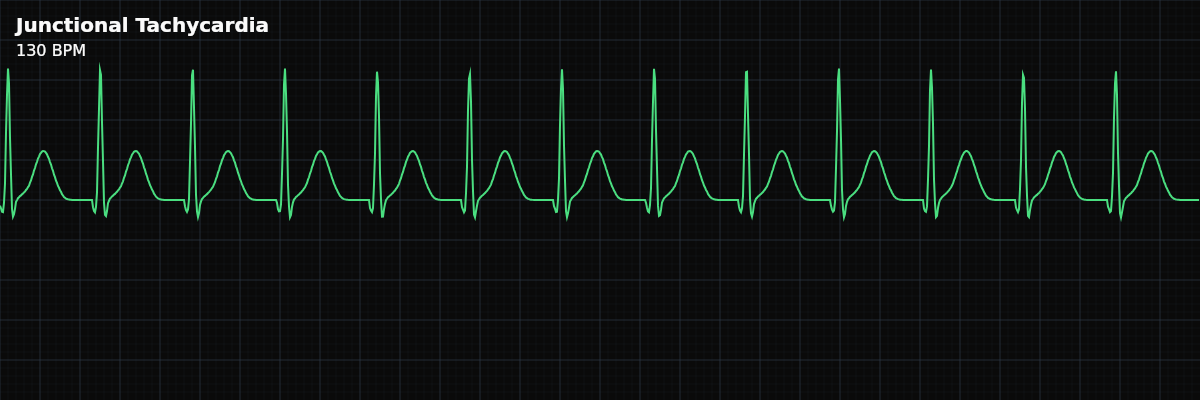
| Rate | > 100 bpm (typically 100–180) |
|---|---|
| Rhythm | Regular |
| P Waves | Absent, retrograde, or hidden |
| PR Interval | Not applicable |
| QRS Duration | < 0.12 s |
Junctional Tachycardia is the AV junction firing at rates it should never reach. The junction's normal rate is 40-60 BPM. In junctional tachycardia, enhanced automaticity pushes the rate above 100 — sometimes to 180 BPM. This is not a backup rhythm filling in for a failed SA node; this is abnormally fast junctional firing that overpowers both the SA node and the normal rate hierarchy.
For monitor technicians, junctional tachycardia is clinically significant for two reasons. First, the rapid rate can compromise cardiac output — especially when combined with the loss of atrial kick. Second, it usually indicates serious underlying pathology: digoxin toxicity, post-cardiac surgery, inferior MI, or myocarditis. This is not a benign rhythm.
What Changed from Normal Sinus Rhythm
Junctional tachycardia replaces the SA node as the dominant pacemaker and drives the ventricles at a rapid rate. The QRS remains narrow (normal His-Purkinje conduction), but the P waves are absent, retrograde, or completely dissociated from the QRS. The rapid rate distinguishes it from the lower junctional rhythms.
Five Criteria: Junctional Tachycardia vs NSR
- Rate: >100 BPM (typically 100-180)
- The fastest junctional rhythm. Post-surgical JET in children can reach 170-260 BPM. In adults, rates are typically 100-180.
- Regularity: Regular
- The enhanced automatic focus fires at a consistent rate. RR intervals are equal. Slight variations may occur with warm-up and cool-down phases.
- P Waves: Absent, retrograde, or dissociated
- P waves may be absent, inverted (retrograde), or marching at a different rate than the QRS (AV dissociation). AV dissociation is particularly common in junctional tachycardia and is an important diagnostic clue.
- PR Interval: Not applicable
- With AV dissociation, P waves bear no consistent relationship to QRS complexes. If retrograde conduction is present, the PR would be very short.
- QRS Complex: Narrow (<0.12s)
- The impulse originates at the AV junction and travels normally through the His-Purkinje system. QRS is identical to sinus-conducted beats.
What Junctional Tachycardia Looks Like on the Strip
On the strip, junctional tachycardia looks like a regular, narrow-complex tachycardia — which makes it look very similar to SVT (AVNRT). The differences are subtle on a short strip. Look for: (1) gradual onset/offset (warm-up/cool-down) rather than abrupt, (2) P waves marching at a different rate than QRS (AV dissociation), and (3) subtle rate variations rather than rock-steady rate.
Junctional Tachycardia vs AVNRT (SVT)
Both are narrow-complex tachycardias, but the mechanism is fundamentally different: **Junctional Tachycardia** — Enhanced automaticity (cells fire too fast). Gradual warm-up at onset, cool-down at termination. AV dissociation is common. Adenosine may slow it but the rhythm typically resumes. **AVNRT (SVT)** — Re-entry circuit through the AV node. Abrupt onset and termination ("like flipping a switch"). 1:1 AV conduction. Adenosine terminates the rhythm by breaking the circuit.
Clinical Context for Monitor Technicians
Junctional tachycardia is most commonly seen in four clinical contexts: digoxin toxicity (classic cause — enhanced automaticity from the drug), post-cardiac surgery (especially in the first 24-72 hours, more common in pediatric patients), inferior MI (ischemia affecting the AV junctional tissue), and inflammatory conditions (myocarditis, pericarditis). It is relatively rare in adults but more common in children post-surgery.
When to Escalate
**Notify immediately:** - New-onset junctional tachycardia (indicates significant underlying pathology) - Rapid rate >150 BPM (hemodynamically significant, especially with loss of atrial kick) - Patient is symptomatic — chest pain, hypotension, altered consciousness - Patient is on digoxin (possible toxicity — clinically urgent) **Notify promptly:** - Post-surgical patient develops junctional tachycardia (expected but needs management) - Rate is increasing (progressive enhancement of automaticity)
Putting It Together
Junctional tachycardia is a regular, narrow-complex tachycardia at >100 BPM with absent, retrograde, or dissociated P waves. It is caused by enhanced automaticity (not re-entry), shows warm-up/cool-down behavior, and is associated with digoxin toxicity, post-cardiac surgery, inferior MI, and myocarditis. AV dissociation is a common finding. Distinguished from AVNRT by its gradual onset/offset and persistence after adenosine.